Essay on How Can the Diagnosis of Lung Cancer Be Improved?
Number of words: 2245
Symptoms and stages of breast cancer
Lung cancer (LC) which is also referred as lung carcinoma, is characterised by a malignant tumour in the lung which is due to the uncontrollable tissue cell growth (1). LC has two main types, small cell lung carcinoma (SCLC) and non-small cell lung carcinoma (NSCLC), and similar to other cancers, LC, is typically initiated by the activation of oncogenes or tumour suppressor genes (1). It is thought that approximately 10-30% of adenocarcinomas are due to K-ras proto-oncogene mutations. Individuals with this condition most often experience symptoms of hemoptysis, chest pains, weight loss and finally shortness of breath (1). Furthermore, LC is also responsible for majority of cancer related deaths (fig. 1).
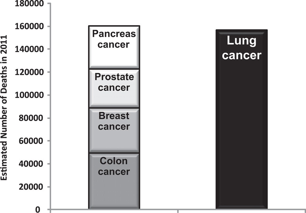
Figure 1 (2)- This graph shows the estimated number of deaths (in 2011) from pancreas, prostate, breast and colon combined. The number of deaths from lung cancer alone is equal to the cumulation of the rest (2).
In regard to the staging process (fig. 2), it refers to the spread from its original location; physicians often use this as a reference for both the treatment plan and prognosis of the cancer (1). NSCLC adopts the TNM classification, this system is effectively based on the size of the primary tumour, spread and potential lymph node involvement (1).
The stages of lung cancer
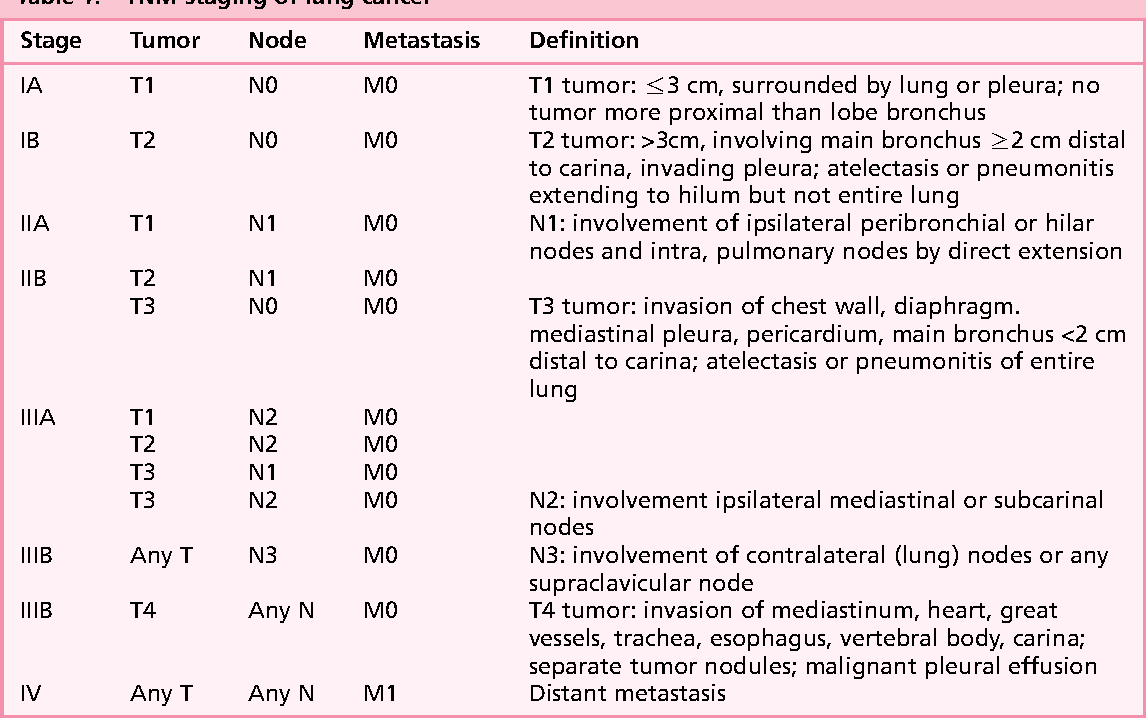
Figure 2 (3): This table shows the different stages of lung cancer and the Tumour, Node, Metastasis classifications (3). As shown, as the stages go up, the cancer gets more aggressive and thus its treatment becomes more difficult (3).
Molecular changes contributing to breast cancer
Furthermore, LC is amongst the leading causes of cancer-related deaths in western nations (fig. 1) (4). Its cause is greatly associated with smoking cigarette (which are mutagens) and 10% of long-term smokers are susceptible (4). The development of this disease is largely due to the build of various molecular abnormalities (fig. 3 and 4). The most common is genomic instability. These alterations are not restricted to one level of gene slicing (4). In NSCLC, the predominant changes that occur in the genome is the loss of the 3p and 9p regions, deletion of chromosomal arm on 5p and mutations of both K-ras and p53 (these have been recognised in later stages) (4). It is important to note that LC can also be caused by chromosomal alternations i.e. amplification and is not solely due to mutations (4). Amplification is mostly seen in chromosome 1q and 3q (4). Genomic instability and alternations are a crucial characteristic of the initiation of LC and it’s the accumulation of the changes that lead to its progression (4).
The loss of chromosome types and their prevalence in nasopharyngeal cancer
| Chromosome type | Prevalence of loss (percentage %) |
| 3p | 75 |
| 11q | 70 |
| 14q | 65 |
| 9q | 60 |
| 13q | 50 |
| 16q | 50 |
Figure 3 (5)- This table shows the chromosomal losses which may occur in primary nasopharyngeal carcinoma. Although this data is not for lung cancer directly, there have been several studies (one of which was conducted by Dr Hui EP in 1999 in Hong Kong titled “Detection of recurrent chromosomal gains and losses in primary nasopharyngeal carcinoma by comparative genomic hybridization.” This study discusses the spread of nasopharyngeal carcinoma to the lungs and this can be used as an early diagnostic factor, as it may be an early indication of development of the cancer to the lung (5).
Chromosomal deletions in small-cell lung cancer and their prevalence
| Chromosome type | Prevalence of loss (percentage %) |
| 3p | 100 |
| 10q | 94 |
| 4q | 86 |
| 5q | 86 |
| 13q | 86 |
| 17q | 86 |
Figure 4 (6)- This table shows details of the molecular changes that occur in lung cancer and specifically deletion of the chromosomes. It can be seen that chromosome 3p accounts 100% of SCLC cases as its prevalence is 100%. Similarly, chromosome 3p possess the highest prevalence loss in nasopharyngeal cancer (fig. 3) (6).
Current methods for breast cancer diagnosis
In the last decade, there has been a lot of progress in the diagnostic methods of LC, having said that, it is still a major medical issue. Some of the current methods of diagnosis include histological assessment (7). Those who are at a higher risk of developing this disease may have more frequent CT scans (8). Clinicians may also conduct other imaging tests like x-rays or sputum tests where they then analyse the sample under a microscope (8). Finally, the patient may also undergo a biopsy (which have many different types) for example, the doctor may wish to test different regions like the throat, lungs or surrounding lymph nodes (8). Furthermore, given that the individual tests return to be positive, the doctor then carries out tests to determine the extent of the spread and stage. This is a crucial step in determining the best possible treatment tailored to that individual, some examples of tests include CT, MRI, bone and PET scans (positron emission tomography) (8). The physician will then determine the stage based on the staging system mentioned above. Although, in most cases these methods are effective, it is important to note that they don’t all have a 100% success rate. Some of disadvantages include, expense (which suggests why clinicians prefer to prevent rather than treat, time and false negatives. As a result, it is vital for diagnostic improvements to be made to prevent progression of the condition and prevent death.
The NHS breast cancer screening programme
Interestingly, there are no current NHS screening programme for LC (9). This is extremely detrimental as around 47,000 cases occur every year with a 5% survival rate (10). Having expressed this, the NHS have arranged to offer regular lung check-ups in certain locations around the UK where the death rates from LC are the highest (starting from Autumn 2019). The NHS argue that they don’t have a national screening programme because the tests have risks (too much radiation that can cause lung damage) and they are also not very cost effective. Another argument is that the screening does not actually saves those who are affected (9). Instead, the NHS aim to conduct national screening programmes for those who are a greater risk of developing the cancer. These include those that smoke and used to smoke, and they aim to use low dose CT scans. Although the NHS do claim that screening has minimal affects in increasing survival rates, there is evidence that suggests that it does save lives based on their risk of LC (9). In certain cases, there can be false positives and therefore further tests like biopsy is needed and these also possess their own risks. Also, false positives may mean that unnecessary treatment is given which again has their own side effects (9). It is essential that a method of diagnosis is developed that has reduced risks and does not have the risk of overdiagnosis (9).
Clinical trials for new diagnostic techniques
There are currently many clinical trials in place for new diagnostic methods, The Roche and Foundation Medicine Collaboration are conducting a system of comprehensive genome profiling which enables specific and tailored treatment for each patient, following the diagnosis (11). Moreover, researchers around worldwide are striving for better ways to diagnose LC; Cancer Research UK are currently looking into biomarkers and proteins that are found in the patients’ blood or urine samples that may give an indication of cancer presence.
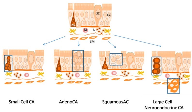
Figure 5 (12)- This image shows the histogenesis of histological types of lung cancer. Histology is a major method of diagnosis (12). Immunohistochemistry which is a method used in histology is used to correctly diagnose LC (12). This is due to the fact that biomarkers cannot solely be used to distinguish the type of LC. This is discussed in a study conducted by Tatiana N. Zamay, titled “Current and Prospective Protein Biomarkers of Lung Cancer,” which explores the different biomarkers used for diagnosis (12)
Moreover, artificial antibodies i.e. aptamers can also be used as recognition elements for tumor markers (12). This method not only allows an easier diagnosis but also earlier given that the cancer returns (9). Another trial currently in place is the detection of circulating tumour DNA (ctDNA): this method has a sensitivity which ranges between 40-93% and specificity of 18-100% (13). As the sensitivity is lower, risks of missing the cancer can be high (this is especially true for early stage cases) (14). Scientists believe that this gives an early diagnosis of NSCLC as a LC tumour can shed many pieces of DNA into the blood (9). Another interesting technique which is currently in trial is the use of sound, researchers have found an interesting difference in the sound produced from people with lung disease in comparison to those who do not have a condition (15).
Conclusion
To conclude, for many years’ lung cancer has been one of the most difficult prognoses and consequently has been a leading cause of death due to cancer worldwide. However, the advancements in research has led to improvement in the diagnosis and medical innovations mainly focus on the early detection of the disease (16). Although early detection is crucial, fast detection is also essential, both of which reduce the morbidity. The future of LC diagnosis holds much promise in restoring the quality and quantity of life for the patients and this will mainly be through a multi-modality approach which include multiple test methods (16).
Works Cited
- contributors w. Lung cancer [Internet]. En.wikipedia.org. 2020 [cited 10 February 2020]. Available from: https://en.wikipedia.org/wiki/Lung_cancer#Staging
- Dela Cruz C, Tanoue L. Lung Cancer: Epidemiology, Etiology, and Prevention [Internet]. PubMed. 2013 [cited 5 March 2020]. Available from: https://www.ncbi.nlm.nih.gov/pmc/articles/PMC3864624/
- Wynants J, Stroobants S, Dooms C, Vansteenkiste J. Staging of Lung Cancer. [Internet]. Semanticscholar.org. 2006 [cited 10 February 2020]. Available from: https://www.semanticscholar.org/paper/Staging-of-Lung-Cancer.-Wynants-Stroobants/23749b44711b125aef5686f1840e4d81bef4f31b/figure/0
- Massion P. The molecular basis of lung cancer: molecular abnormalities and therapeutic implications. pubmed. 2020.
- Hui AB e. Detection of recurrent chromosomal gains and losses in primary nasopharyngeal carcinoma by comparative genomic hybridisation. – PubMed – NCBI [Internet]. Ncbi.nlm.nih.gov. 1999 [cited 5 March 2020]. Available from: https://www.ncbi.nlm.nih.gov/pubmed/10404061
- Petersen I, Langreck H. Small-cell lung cancer is characterized by a high incidence of deletions on chromosomes 3p, 4q, 5q, 10q, 13q and 17p. [Internet]. PubMed. 1997 [cited 5 March 2020]. Available from: https://www.ncbi.nlm.nih.gov/pmc/articles/PMC2222682
- 7.Hammerschmidt, Stefan, and Hubert Wirtz. “Lung cancer: current diagnosis and treatment.” Deutsches Arzteblatt international vol. 106,49 (2009): 809-18; quiz 819-20. doi:10.3238/arztebl.2009.0809
- contributors m. Lung cancer – Diagnosis and treatment – Mayo Clinic [Internet]. Mayoclinic.org. 2020 [cited 10 February 2020]. Available from: https://www.mayoclinic.org/diseases-conditions/lung-cancer/diagnosis-treatment/drc-20374627
- contributors c. Screening | Lung cancer | Cancer Research UK [Internet]. Cancerresearchuk.org. 2020 [cited 10 February 2020]. Available from: https://www.cancerresearchuk.org/about-cancer/lung-cancer/getting-diagnosed/screening
- contributors c. Lung cancer statistics [Internet]. Cancer Research UK. 2020 [cited 10 February 2020]. Available from: https://www.cancerresearchuk.org/health-professional/cancer-statistics/statistics-by-cancer-type/lung-cancer
- contributors r. Roche Foundation Medicine | What We Do [Internet]. Rochefoundationmedicine.com. 2020 [cited 10 February 2020]. Available from: https://www.rochefoundationmedicine.com/home/what-we-do.html
- Zamay T, Zamay G. Current and Prospective Protein Biomarkers of Lung Cancer [Internet]. PubMed. 2017 [cited 5 March 2020]. Available from: https://www.ncbi.nlm.nih.gov/pmc/articles/PMC5704173/
- Passiglia F, Rizzo S. The diagnostic accuracy of circulating tumor DNA for the detection of EGFR-T790M mutation in NSCLC: a systematic review and meta-analysis [Internet]. Nature.com. 2018 [cited 5 March 2020]. Available from: https://www.nature.com/articles/s41598-018-30780-4
- Fiala C, Diamandis E. Utility of circulating tumor DNA in cancer diagnostics with emphasis on early detection [Internet]. PubMed. 2018 [cited 5 March 2020]. Available from: https://www.ncbi.nlm.nih.gov/pmc/articles/PMC6167864/
- contributors c. Research into lung cancer early diagnosis and screening | Cancer Research UK [Internet]. Cancerresearchuk.org. 2018 [cited 10 February 2020]. Available from: https://www.cancerresearchuk.org/about-cancer/lung-cancer/research-clinical-trials/research-diagnosis-screening
- Blandin Knight S, Crosbie PA, Balata H, Chudziak J, Hussell T, Dive C. Progress and prospects of early detection in lung cancer. Open Biol. 2017;7(9):170070. doi:10.1098/rsob.170070